Leiomyosarcoma Treatment in Pune
Sancheti Hospital’s Ortho Onco Department provides expert treatment for leiomyosarcoma with advanced surgical care.
Overview
Leiomyosarcoma (LMS) is a rare type of cancer that develops in the smooth muscles of the body. Unlike the muscles we can control voluntarily, smooth muscles work automatically to help with important body functions like digestion, blood flow, and organ operation. Since these muscles are found throughout the body, LMS can develop in almost any organ, making it a type of soft tissue sarcoma.
This cancer is particularly challenging because it grows aggressively and unpredictably. Tumors can double in size in just one month and spread quickly to other parts of the body. However, when LMS is caught early and treated before it spreads, the outlook is much better.
Symptoms
LMS symptoms vary greatly depending on where the tumor grows and how large it becomes. In early stages, many people experience no symptoms at all, which makes early detection difficult. As the tumor grows, several signs may appear:
General symptoms include:
- A firm, usually painless lump under the skin
- Unexplained tiredness and fatigue
- Fever without obvious cause
- Unintentional weight loss
- Pain or discomfort in the affected area
- Nausea and vomiting
- Abdominal bloating (if the tumor is in the abdomen)
Location-specific symptoms:
- Digestive system: Stomach pain, black stools (indicating internal bleeding), loss of appetite, and vomiting blood
- Uterine leiomyosarcoma: Abnormal vaginal bleeding unrelated to menstruation, unusual discharge, frequent urination, or pelvic pain and pressure
Causes
The exact causes of Leiomyosarcoma remain unknown to medical researchers. However, scientists believe it results from genetic changes within cells that cause uncontrolled growth and tumor formation. These genetic changes can happen spontaneously or may be inherited from family members.
Research has identified connections between LMS and certain genetic conditions that may increase a person’s risk:
- Hereditary retinoblastoma
- Li-Fraumeni syndrome
- Neurofibromatosis type 1 (NF1)
- Tuberous sclerosis
- Nevoid basal cell carcinoma syndrome (Gorlin syndrome)
- Gardner syndrome
- Werner syndrome
Despite these genetic links, most LMS cases are not directly connected to family history. Scientists continue studying this cancer to better understand how it develops and spreads.
Risk Factors
While the specific causes of Leiomyosarcoma are often unclear, several factors may increase a person’s risk of developing this condition:
Age: LMS is more common in adults, especially those over 50 years old. Uterine leiomyosarcoma typically affects perimenopausal women around their fifth decade of life.
Radiation exposure: Previous radiation therapy for other types of cancer significantly increases the risk of developing soft tissue sarcomas, including LMS, years later.
Chemical exposure: Contact with certain harmful chemicals like dioxins (found in pesticides and paper manufacturing) and vinyl chloride (used in plastic production) has been linked to increased risk. Some weedkillers may also contribute.
Medical treatments: Tamoxifen therapy for breast cancer has been associated with uterine leiomyosarcoma in some cases.
Reproductive factors: Never having been pregnant (nulliparity) and hormone use during menopause may increase risk for uterine leiomyosarcoma.
Obesity: Some studies suggest that being overweight may increase the risk of developing uterine leiomyosarcoma.
Having one or more risk factors doesn’t guarantee someone will develop LMS. Many people with LMS have no identifiable risk factors at all.
Diagnosis
Accurate diagnosis of Leiomyosarcoma is critical because of its aggressive nature and impact on prognosis. The diagnostic process typically involves several important steps:
Medical history and physical examination: Your doctor will ask about your symptoms and medical background, then perform a physical exam to check for lumps or other concerning signs.
Imaging tests: These are essential for seeing the tumor, determining its size and location, and checking if it has spread to other parts of the body:
- CT scans: Provide detailed cross-sectional images
- MRI: Uses magnets and radio waves for highly detailed pictures
- PET scans: Detect cancer cells based on their activity level
- Chest and abdominal CT scans are typically required since LMS often spreads to the lungs and liver
Biopsy: This is the most important step for confirming LMS diagnosis. A small tissue sample is taken from the tumor and examined under a microscope by a pathologist to determine if cancer cells are present and identify the specific type.
Given the rarity and complexity of LMS, it’s highly recommended to seek diagnosis from a team of doctors experienced in soft tissue sarcomas. Expert review by a specialized pathologist is often necessary to ensure accuracy.
Types
Leiomyosarcoma can be classified into three main types based on where it originates:
Somatic soft tissue LMS: This is the most common form, affecting connective tissues throughout the body. Uterine leiomyosarcoma is the most frequent example, as the uterus contains large amounts of smooth muscle. Other common locations include the abdomen, arms, legs, and trunk.
Cutaneous or subcutaneous LMS: This type affects the small muscles in the skin and eyes that cause goosebumps and control pupil size.
Vascular LMS: This is the rarest form, developing in major blood vessels like the pulmonary arteries, inferior vena cava, or peripheral arteries.
Stages
Staging describes how far the cancer has spread and helps guide treatment decisions. The staging system used depends on where the LMS originates:
Uterine leiomyosarcoma uses the FIGO staging system:
- Stage I: Tumor is limited to the uterus
- Stage II: Cancer extends within the pelvis
- Stage III: Cancer invades abdominal tissues
- Stage IV: Cancer involves distant organs
Extrauterine leiomyosarcoma uses the AJCC staging system, which considers:
- Tumor size: The largest dimension of the tumor
- Lymph node involvement: Whether regional lymph nodes contain cancer
- Metastasis: Presence of distant spread
Tumor grading also plays a crucial role, reflecting how aggressive the cancer is:
- Grade 1: Slow-growing, less aggressive
- Grade 2: Moderately aggressive
- Grade 3: Fast-growing, highly aggressive
Most LMS cases are diagnosed as high-grade (Grade 2 or 3), indicating more aggressive behavior.
Treatment
Treatment for Leiomyosarcoma is highly individualized based on the tumor’s location, size, grade, spread, and the patient’s overall health. A team approach involving multiple specialists is essential for the best outcomes.
Surgery is the most important treatment for localized LMS and offers the best chance for cure when the entire tumor can be removed. The goal is complete removal with clear margins (no cancer cells at the edges).
- Extremity or trunk LMS: Surgeons work to remove the tumor while preserving important nerves, blood vessels, and limb function
- Retroperitoneal LMS: These tumors can be challenging to remove completely due to their location near vital organs
- Uterine leiomyosarcoma: Standard treatment involves hysterectomy (removal of the uterus) and sometimes removal of ovaries
Radiation therapy uses high-energy X-rays to kill cancer cells and is often used alongside surgery. It can be given before surgery to shrink the tumor or after surgery to eliminate remaining cancer cells. The timing depends on the tumor’s location and other factors.
Chemotherapy uses medications to kill cancer cells throughout the body. It’s typically recommended for large tumors or when cancer has spread to other parts of the body. LMS responds moderately well to chemotherapy.
Common chemotherapy drugs include:
- Doxorubicin (often combined with ifosfamide)
- Gemcitabine and docetaxel (particularly effective for uterine leiomyosarcoma)
- Trabectedin for advanced cases
- Pazopanib for metastatic disease
Targeted therapy drugs attack specific features of cancer cells, often with fewer side effects than traditional chemotherapy. Examples include pazopanib and anlotinib.
Immunotherapy helps the body’s immune system fight cancer. While it hasn’t shown strong results as a single treatment for LMS, combinations with other treatments are being studied and show promise.
Rehabilitation
After Leiomyosarcoma treatment, rehabilitation plays a vital role in recovery and improving quality of life. Recovery time varies significantly based on the tumor’s size, location, and treatments received.
Rehabilitation may include:
- Physical therapy to regain strength and mobility
- Occupational therapy to help with daily activities
- Pain management strategies
- Nutritional support
- Psychological counseling
Regular follow-up appointments with the oncology team are essential to monitor for recurrence and manage any long-term side effects from treatment.
Complications
Leiomyosarcoma and its treatment can lead to various complications:
Tumor-related complications:
- Large tumors may compress surrounding organs, causing pain or dysfunction
- Early spread to lungs, liver, or bones is common due to the cancer’s aggressive nature
Treatment-related side effects:
- Chemotherapy: Nausea, vomiting, hair loss, fatigue, anemia, and fertility problems
- Radiation therapy: Skin irritation, pain, swelling, and long-term tissue changes
- Surgery: Wound healing issues, infection, and functional limitations
- Immunotherapy: Immune-related side effects that require careful monitoring
Prevention
Currently, there is no known way to prevent Leiomyosarcoma. However, people can reduce their overall risk of developing certain types of sarcoma by:
- Avoiding unnecessary radiation exposure
- Limiting contact with harmful industrial chemicals when possible
- Following safety guidelines in workplaces that use chemicals
- Maintaining a healthy lifestyle with regular exercise and balanced nutrition
Despite these measures, many LMS cases occur without identifiable risk factors. The focus remains on early detection and prompt, specialized treatment to improve outcomes.
Living With Leiomyosarcoma
Receiving a diagnosis of Leiomyosarcoma can be overwhelming, affecting both physical and emotional well-being. It’s important to prioritize comprehensive care and support throughout the journey.
- Maintaining a balanced diet and staying physically active as tolerated
- Getting adequate sleep and managing stress effectively
- Following all medical appointments and treatment plans
- Communicating openly with healthcare providers about concerns
Key Takeaways
- Leiomyosarcoma is a rare, aggressive smooth muscle cancer that can develop anywhere in the body
- Early symptoms are often absent, making regular medical checkups important
- Diagnosis requires specialized imaging and biopsy by experienced pathologists
- Surgery remains the primary treatment, with radiation therapy and chemotherapy as important additions
- Prognosis depends heavily on early detection, tumor size, location, and extent of spread
- Comprehensive care includes physical rehabilitation and emotional support
- Research continues into new treatments including targeted therapy and immunotherapy
At Sancheti Hospital, we understand the complexity and challenges of treating rare cancers like Leiomyosarcoma. Our multidisciplinary team of experienced oncologists, surgeons, and support staff is dedicated to providing comprehensive, personalized care for each patient.
We offer state-of-the-art diagnostic facilities, advanced treatment options, and compassionate support throughout the entire treatment journey. Our hospital is equipped with the latest technology for surgery, radiation therapy, and chemotherapy, ensuring that patients receive the highest standard of care.
We also provide rehabilitation services and psychological support to help patients and their families navigate this challenging diagnosis. For those facing Leiomyosarcoma, Sancheti Hospital is committed to delivering expert care with dignity and hope.
Meet Our Ortho Onco Specialist
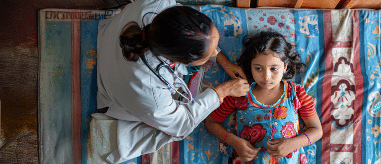
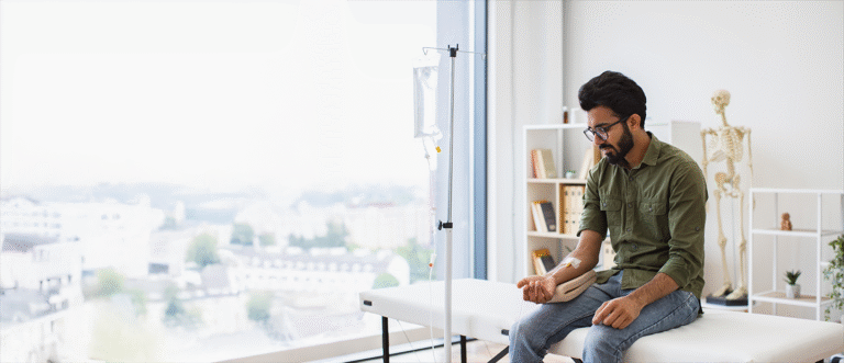
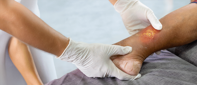
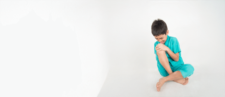
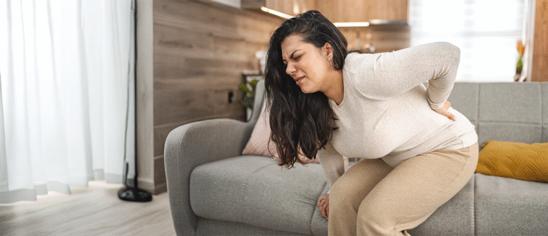
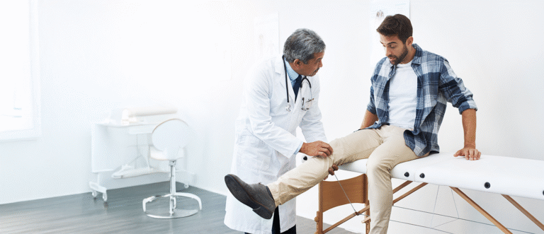
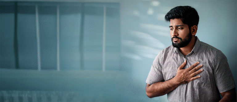
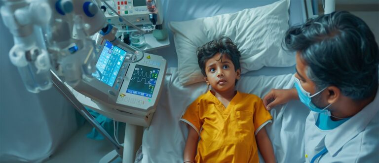
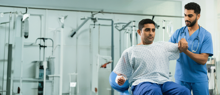
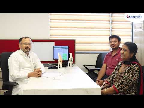
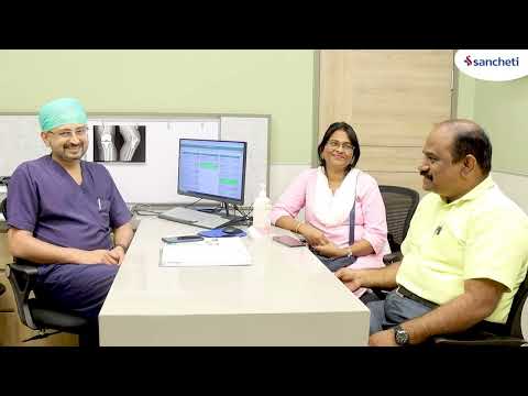
Patient Stories & Experiences
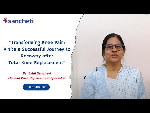
The pain in my left knee left me feeling helpless for years. After my treatment here, I can finally say I'm pain-free.
Vinita Singh
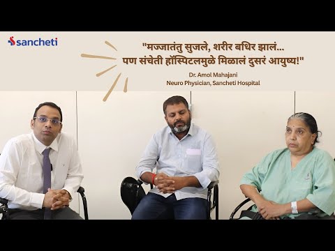
The nerves were swollen, and the body went numb. But thanks to Sancheti Hospital, I got a second life!
Parvati
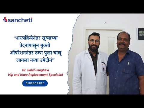
I finally could walk again, a relief I've only felt after the hip pain surgery. I thank the doctors at Sancheti Hospital for their help.
Balaji Kharat
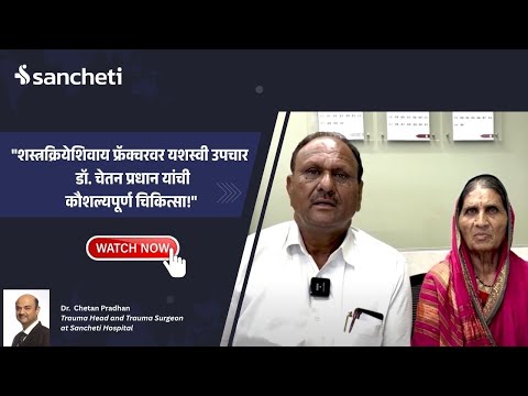
I'm a police officer, and I'm extremely thankful to Sancheti Hospital for treating my fracture without surgery.
Shantilal
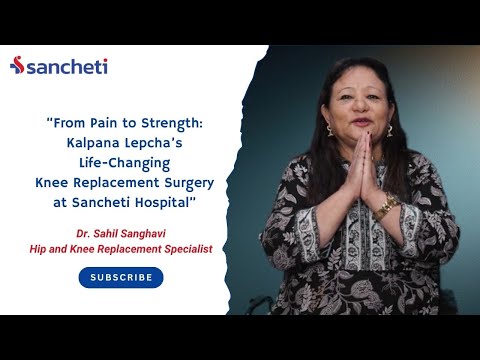
My life has completely changed after the knee replacement surgery at Sancheti Hospital. It's like I can finally live again!
Kalpana Lepcha
The knee pain I've carried for years finally went away with the help of Sancheti Hospital.
Karuna
I can't believe that I get to finally live a normal and happy life, all thanks to the knee surgery I had at Sancheti Hospital.
Kishore Bhosle
Other Conditions
Frequently Asked Questions
What are the odds of getting Leiomyosarcoma?
Leiomyosarcoma is very rare, affecting approximately 1 to 1.4 cases per 100,000 people annually. It’s slightly more common in women than men and primarily affects individuals over 50 years old.
What are the early symptoms of Leiomyosarcoma?
Early symptoms are often absent or very subtle. As the tumor grows, you might notice an unexplained lump, pain, fatigue, weight loss, or digestive issues. Uterine leiomyosarcoma may cause abnormal vaginal bleeding.
Does Leiomyosarcoma run in families?
Most LMS cases occur sporadically without family history. However, it can be associated with certain rare inherited genetic conditions like Li-Fraumeni syndrome and hereditary retinoblastoma.
Can you live a long life with Leiomyosarcoma?
The possibility of long-term survival depends on several factors including tumor size, location, grade, and whether it has spread. Early detection and complete surgical removal significantly improve the prognosis.
What is the survival rate for Leiomyosarcoma?
Five-year survival rates vary by stage: localized disease (60-63%), regional spread (36-37%), and distant spread (12-14%). These are estimates, and individual outcomes can vary significantly.
What is the treatment for uterine leiomyosarcoma?
The primary treatment is surgery (hysterectomy) to remove the uterus and sometimes ovaries. Chemotherapy and radiation therapy may be added depending on the stage and risk factors.
Can leiomyoma become leiomyosarcoma?
It’s extremely rare, but benign uterine fibroids (leiomyomas) can very occasionally transform into leiomyosarcoma. This is why rapidly growing masses, especially after menopause, should be evaluated promptly.
Home
Patient Login
International patients
Contact Us
Emergency
Download Reports

