Home
Home PG College
 Patient Login
 Sancheti Clinic
 Contact Us
 Download Reports
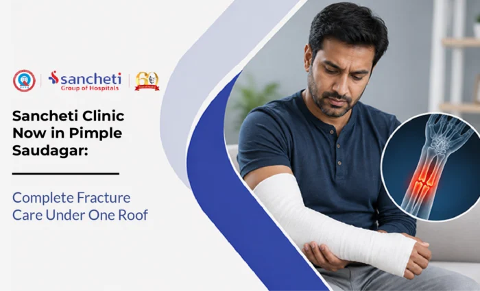

Sancheti Clinic Now in Pimple Saudagar: Complete Fracture Care Under One Roof
A bone fracture is a complete or partial break in bone continuity caused by force that exceeds the bone’s structural strength. For residents of Pimpri-Chinchwad, fractures are one of the most common orthopaedic emergencies, driven by the area’s active construction and manufacturing workforce, and growing sports participation across all age groups.
Sancheti Hospital’s 24/7 Emergency and Trauma Department in Pune provides round-the-clock specialist fracture care for patients across the region. The centre manages everything from straightforward forearm fractures to complex pelvic trauma and neglected non-union cases. This guide explains fracture types, treatment decisions, and what recovery looks like at each stage.
What Is Fracture Treatment?
Fracture treatment is the process of restoring a broken bone to anatomical alignment, stabilising it so healing can occur, and rehabilitating the surrounding soft tissue to restore full function. The treatment approach depends on several factors working together: which bone is fractured, whether the fracture is open or closed, how much the bone fragments have shifted from their normal position, the patient’s age and bone quality, and the functional demands the bone will need to meet after healing.
The goal is not simply for the bone to join together. It is for the bone to heal in the correct position, with normal length and rotation, so that the limb functions well long-term. A fracture that heals in a malunited position can cause permanent pain, reduced mobility, and early joint degeneration in adjacent joints. This distinction between bone union and functional recovery shapes every decision in fracture management.
Who Is Susceptible to Fractures
Fractures affect every age group, but the pattern of injury shifts significantly across the lifespan. In India, vehicular accidents account for nearly half of all fracture presentations at tertiary centres, with the 18-50 age group bearing the heaviest burden (International Journal of Research in Orthopedics, Indian Fracture Epidemiology). In PCMC specifically, two-wheeler accidents on the Pune-Mumbai highway corridor and inner industrial roads are a primary source of tibial shaft fractures, femoral fractures, and forearm injuries in working-age men.
Falls account for a large proportion of fractures in older adults, where osteoporosis reduces bone strength to the point where a low-energy fall from standing height can fracture the hip, wrist, or vertebra. At the other end of the age spectrum, children fracture forearm bones and growth plates in sports and playground accidents. A dedicated fracture doctor in PCMC assesses these presentations very differently, since management varies substantially by age and bone quality.
Types of Fractures
Fractures are classified by whether the skin is broken, how much the fragments have moved, and which bone is involved. These distinctions directly determine the treatment pathway.
| Fracture Type | Key Characteristic | Typical Cause in Working-Age Adults |
|---|---|---|
| Closed fracture | Bone broken, skin intact; most common type | Low- to high-energy falls, road accidents, sports collisions |
| Open (compound) fracture | Bone pierces or is exposed through the skin; surgical emergency | High-velocity road accidents; falls from height in industrial settings |
| Displaced fracture | Fragments shifted from normal alignment; usually requires reduction | High-energy impacts; falls on outstretched hand |
| Comminuted fracture | Bone broken into three or more fragments | High-velocity trauma; direct crushing force |
| Stress fracture | Hairline crack from repetitive loading, not a single impact | Runners, military recruits, construction workers with high step count |
| Fragility (osteoporotic) fracture | Low-energy fracture in bone weakened by osteoporosis | Minor falls in adults over 55; wrist, hip, and vertebra most common |
Symptoms and When to Seek Emergency Care
Some fractures are unmistakable: a bone visibly deformed after a road accident, an ankle that collapses under weight after a sports injury. Others are less obvious, particularly stress fractures and undisplaced fragility fractures in older adults, where the primary symptom is localised pain that worsens with activity and persists at rest. In either case, any bone or joint that is significantly painful after a traumatic event or impact needs imaging before being dismissed as a sprain or bruise.
Seek orthopaedic emergency care immediately if any of these are present after an injury: visible bone through the skin; significant deformity or abnormal angulation of a limb; numbness, tingling, or absent pulse below the injury site, which suggest nerve or vascular compromise; inability to bear weight after a lower limb injury; severe pain that does not ease with rest; or joint swelling that appears within minutes of an injury rather than hours. These presentations require assessment at a facility equipped for emergency fracture surgery, not a clinic.
How Fractures Are Diagnosed
Fracture diagnosis is primarily a clinical and imaging exercise. The orthopaedic surgeon assesses the mechanism of injury, the location and pattern of pain, swelling and deformity, and the neurovascular status of the limb below the fracture before any imaging is ordered. This clinical assessment determines the urgency and the type of imaging needed.
- X-ray: the standard first investigation for any suspected fracture; two views at right angles to the injury site are essential, since a fracture visible on one plane may be missed on a single view.
- CT scan: required for complex fractures involving joint surfaces (wrist, ankle, tibial plateau, acetabulum) and for surgical planning; shows three-dimensional fragment arrangement that X-ray cannot.
- MRI: used for stress fractures not visible on X-ray, and for bone marrow oedema in suspected fragility fractures of the hip or spine before collapse is radiologically apparent.
- Neurovascular assessment: pulse check, capillary refill, and sensation testing below the fracture level at every assessment; a limb with absent distal pulse is a vascular emergency.
Fracture Treatment: Conservative and Surgical Options
The decision between conservative and surgical fracture management depends on fracture stability, displacement, the bone involved, and what is needed for the patient to regain function.
Conservative management works reliably for undisplaced or minimally displaced fractures in bones that a cast or splint can adequately immobilise.
Surgical fixation is indicated when a fracture cannot be held in acceptable position by a cast, when joint involvement makes anatomical reduction essential to prevent arthritis, or when the patient’s functional needs require early weight-bearing. Sancheti Hospital’s Orthopaedic Trauma and Fracture Fixation team performs the full range of modern fixation techniques.
- Intramedullary nailing: a metal rod is inserted through a small incision into the bone canal
- Open reduction and internal fixation (ORIF) with plates and screws: for fractures involving joint surfaces, wrist fractures, forearm fractures, and ankle fractures
- External fixation: used as a temporary stabiliser for open fractures and severely contaminated wounds
- Hip hemiarthroplasty: for displaced neck-of-femur fractures in older adults
Recovery After Fracture Treatment
Bone healing follows a biological timeline that cannot be shortened significantly by any intervention. Rehabilitation ensures the surrounding muscles, joints, and functional patterns are restored optimally alongside that healing, so the patient returns to full use of the limb as soon as the bone allows.
| Stage | Timeframe | Focus of Recovery |
|---|---|---|
| Acute protection | Weeks 1-3 | Immobilisation or post-surgical wound healing; swelling control; adjacent joint gentle movement to prevent stiffness |
| Early rehabilitation | Weeks 3-8 | Physiotherapy begins; range of motion exercises; progressive weight-bearing as bone callus forms; muscle activation |
| Progressive loading | Weeks 6-16 | Increasing load through the healing bone; strength building in the limb; gait correction after lower limb fractures |
| Return to full activity | 3-6 months (varies by bone and fixation) | Return to work, driving, sport; confirmed by clinical exam and X-ray showing union |
A physiotherapist guides each stage of recovery. The most common cause of a poor functional outcome after a well-fixed fracture is inadequate rehabilitation, not the surgery itself.
Prevention and Risk Reduction For Fractures
While high-energy fractures from road accidents cannot always be prevented, a significant proportion of fractures in every age group are the result of modifiable risks. Addressing these directly reduces both the likelihood and severity of fractures.
- Two-wheeler road safety: wearing a helmet and appropriate protective riding gear substantially reduces both fracture severity and the risk of head injury in an accident.
- Workplace fall prevention: scaffolding standards, appropriate footwear on industrial floors, and anti-fatigue matting reduce fall-from-height and slip fractures in the manufacturing workforce.
- Osteoporosis management: bone density assessment, vitamin D and calcium adequacy, and bisphosphonate therapy where indicated significantly reduce fragility fracture risk in adults over 50.
- Strength and balance training: resistance exercise improves bone density over time; balance training specifically reduces fall risk in older adults, the population most vulnerable to hip and wrist fragility fractures.
- Appropriate sports equipment and technique: wrist guards for skating and cycling, proper footwear for running, and sport-specific warm-up protocols reduce stress fractures and impact fractures.
Key Takeaways
- Fracture treatment in PCMC aims to restore the bone’s normal alignment and function, ensuring long-term mobility rather than simply allowing the bone to heal.
- Early assessment is essential because the type, location, and severity of a fracture determine whether it can be managed with immobilisation or requires surgical fixation.
- Not all fractures are obvious, and persistent pain, inability to bear weight, or swelling after an injury should always be evaluated with appropriate imaging.
- Rehabilitation is just as important as fracture healing, with physiotherapy helping restore strength, joint movement, balance, and confidence once the bone is stable.
- Recovery follows the body’s natural healing process, and returning to work, driving, or sports should be guided by clinical assessment rather than time alone.
- Fracture risk varies across different age groups, making injury prevention, workplace safety, bone health, and fall prevention important strategies for reducing serious fractures.
- Prompt specialist care for complex, open, or displaced fractures improves healing, lowers the risk of complications, and supports better long-term functional outcomes.
- Patients seeking a bone fracture hospital in Pimpri or an orthopaedic emergency near Pimpri-Chinchwad in Pune can access round-the-clock trauma care at Sancheti Hospital’s Kamalnayan Bajaj Trauma Centre.
Frequently Asked Questions (FAQs)
Q1. How do I know if I have a fracture or just a bad sprain?
The honest answer is that you often cannot tell reliably without an X-ray, and neither can a doctor from examination alone. Significant swelling, bruising, and point tenderness over a bone after an injury are all present in both severe sprains and undisplaced fractures, and the clinical picture overlaps substantially. The risks of missing a fracture, including malunion, chronic pain, and joint instability if inadequately treated, are significantly higher than the inconvenience of an X-ray that turns out to show no fracture. Any injury where you cannot bear weight comfortably, where there is focal bone tenderness, or where swelling appeared within minutes rather than hours should be assessed at an orthopaedic facility with imaging available. Do not rely on the absence of visible deformity to rule out a fracture.
Q2. Does every fracture need surgery?
No. Approximately 20% of fractures at Indian tertiary centres are managed conservatively, and many more could be managed non-operatively if the patient’s functional demands allow. Undisplaced fractures, fractures in bones that immobilise well in a cast, and stress fractures in compliant patients often heal completely without any surgical intervention. The decision for surgery is driven by whether the fracture can be held in an acceptable position by external means, whether joint involvement requires precise anatomical reduction, and whether the patient’s life or work demands make early weight-bearing essential. A fracture that surgery could fix is not necessarily a fracture that needs surgery if conservative management will produce an equivalent functional outcome. The orthopaedic surgeon’s job is to make that judgement for each specific case, not to default to the most technically complex option available.
Q3. My elderly parent fell and is in pain but can move the hip. Is it still a fracture?
Possibly, yes. Undisplaced hip fractures in elderly patients with osteoporotic bone can sometimes allow limited weight-bearing immediately after the fracture, because the fragments are still in contact and the muscle forces of the hip temporarily hold the position. This partial mobility is frequently misinterpreted by families as evidence that no fracture has occurred, delaying the imaging that would show it. An elderly adult who falls and has persistent groin pain, inner thigh pain, or difficulty bearing weight after a fall needs same-day imaging of the hip regardless of whether they can move the limb. Delayed diagnosis of an undisplaced hip fracture allows it to displace fully, converting what might have been a simpler internal fixation procedure into a more complex reconstruction. Time matters specifically in this age group.
Q4. How long will I be in a cast, and when can I return to work?
Cast duration depends on the bone fractured, the type of fracture, the patient’s age, and bone quality. As a general guide: wrist fractures are typically casted for four to six weeks; ankle fractures for six to eight weeks; tibial shaft fractures for eight to twelve weeks if managed non-operatively. Children heal faster than adults, often at two-thirds the adult timeline for equivalent fractures. Return to work depends on the physical demands of the job. A desk-based professional can often return within weeks of a hand or foot fracture with appropriate cast protection; a manual worker in construction or manufacturing may need three to four months off after a lower limb fracture requiring surgery. The fracture doctor and physiotherapist will give a specific return-to-work timeline based on your X-ray findings and the nature of your work.
Q5. What is a non-union fracture and can it be treated?
Non-union is the failure of a fracture to heal within the expected biological timeframe, typically defined as no evidence of healing progression on X-ray over three consecutive months. It is more common in open fractures, in fractures with poor initial stabilisation, in smokers, in diabetic patients with impaired circulation, and in cases where the bone fragments are too widely separated to allow bridging callus to form. Non-union does not mean the fracture is untreatable. Most non-unions respond to surgical intervention: debridement of the non-healing tissue at the fracture site, renewed fixation with more rigid hardware, and bone grafting to stimulate a new healing response. Sancheti Hospital’s trauma team specifically manages neglected and ununited fractures as a subspecialty, including complex reconstruction cases that have failed treatment elsewhere.